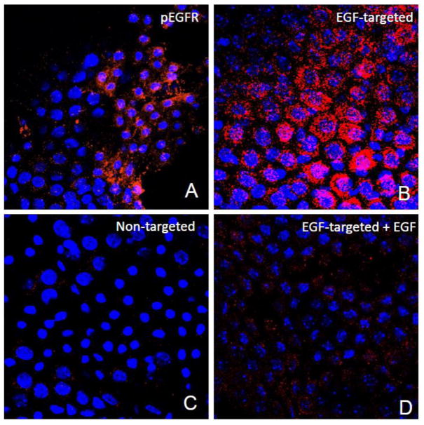
Figure 4. Internalization of EGF-targeted phage is specific in CP explants.
Choroid plexus were dissected from the lateral and 4th ventricle and incubated with EGF-targeted (Panels A, B &D) or non targeted phage (Panel C) at a concentration of 1×10¹²cfu/ml for 2h at 37C. To remove any particles on the cell surface, the explants were washed with PBS containing Tween 20, fixed and then immunostained with antibodies against phosphorylated EGFR (Panel A) or M13 phage (Panels B-D). Binding of primary antibodies was detected using 594 Alexa-donkey anti goat (Panel A) or 488 Alexa-goat anti rabbit antibodies (Panels B-D). Positive immunostaining for the phosphorylated form of the EGFR receptor was observed in the choroid epithelial cells after incubation with specific antibodies (Panel A). Internalization of EGF-phage is observed in the cytoplasm of the epithelial cells (Panel B). In contrast, no immunoreactivity is observed when the explants are incubated with untargeted phage (Panel C). Internalization of EGF-targeted phage is blocked in the presence of an excess of EGF (Panel D). Tissue explants were counterstained with DAPI to visualize the cell nuclei. pEGFR: Red; M13 phage: Red; Cell nuclei: Blue.

