
Figure 1.
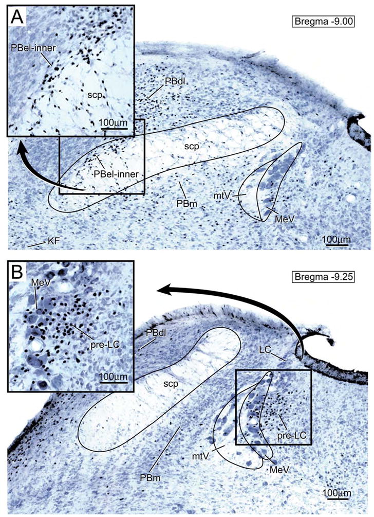
Transverse brainstem section showing the distribution of FoxP2-ir neurons in the parabrachial nucleus (PB). A. The insert shown in the upper left hand corner presents an enlargement of the FoxP2-ir neurons in the external lateral parabrachial subnucleus- inner division (PBel-inner). Other abbreviations: KF= Kölliker-Fuse nucleus; PBdl= dorsal lateral parabrachial subnucleus; PBm= medial parabrachial subnucleus; MeV = mesencephalic nucleus of the trigeminal nerve; mtV = tract of the mesencephalic trigeminal nucleus; scp= superior cerebellar peduncle.
B. Transverse brainstem section showing the pre-locus coeruleus nucleus (pre-LC). This nucleus lies immediately rostral to the locus coeruleus within the lateral part of the periventricular gray matter. As shown in the enlargement in the upper left hand corner, the FoxP2-ir neurons are concentrated in the zone medial to the mesencephalic nucleus of the trigeminal nerve (MeV), but some are interspersed among the MeV neurons..