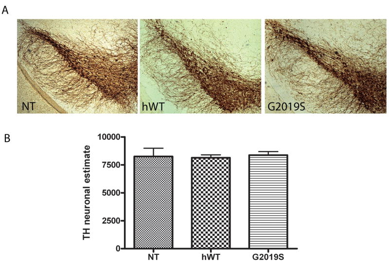
Figure 3. Stereological estimates of TH neurons in the substantia nigra reveal no differences between NT, hWT and G2019S mice.
Counting was performed using the optical fractionator probe in tissue processed for TH staining from hWT, G2019S and NT mice aged 22–24 months. (A) TH immunostaining in the nigra of NT, hWT and G2019S mice (B) Neuronal estimates in NT, hWT and G2019S mice. Data is plotted as mean ± SEM.

