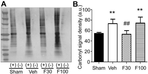
Fig. 6.
Effect of fenofibrate on protein oxidation in the ischemic hemisphere at 30 minutes after reperfusion following 60 minutes of MCAO. Mice were treated with vehicle (Veh) or fenofibrate [30 mg/kg (F30) or 100 mg/kg (F100)] for 7 days and subjected to MCAO at 1 hour after the last drug treatment. Non-drug treated animals that were subjected to sham surgery served as sham control. (A) Western blot analysis showing derivitized protein carbonyls. Brain lysates (10 μg protein) were treated with (+) or without (−) 2,4-dinitrophenylhydrazine and separated by SDS-PAGE. Left end lane was loaded protein marker. (B) bargraph showing density of whole lane of each sample. Error bars, SD. **, p < 0.01 compared to Sham group and ##, p < 0.01 compared to Veh group by ANOVA followed by Scheffe (n = 6 or 7 in each group).

