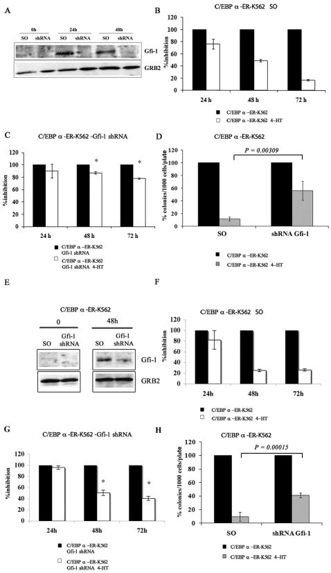
Figure 3. Expression of Gfi-1 is required for C/EBPα-dependent proliferation inhibition in K562 cells.
(A and E) Western blot shows Gfi-1 expression in 4-HT-treated scramble or Gfi-1 shRNA-transduced or transfected C/EBPα-ER/K562 cells; histograms show: proliferation of 4-HT-treated scramble (B and F) or Gfi-1 shRNA (C and G)-transduced or transfected C/EBPα-ER-K562 cells and methylcellulose colony formation of 4-HT-treated scramble or Gfi-1 shRNA-transduced or transfected C/EBPα-ER-K562 cells (D and H). Expression of Gfi-1 was detected by anti-Gfi-1 goat polyclonal antibody (N-20; Santa Cruz Biotechnology). For cell count assays, cells were seeded at 105cells/ml; for colony formation assays, 1,000 cells/plate were seeded. Data (expressed as % inhibition of 4-HT-treated vs untreated cells) are represented as the mean ± SEM of 2 (B-D) or 3 (F-H) independent experiments. * denotes that the differences in cell counts between Gfi-1 and scramble shRNA K562 cells (compare panels C,G and B,F) are statistically significant (p= 0.001 and 0.002, respectively ).

