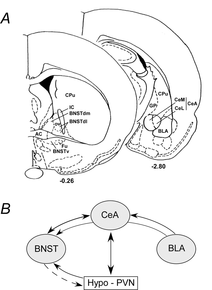
Figure 2. Schematic of extended amygdala areas and interactive pathways.
A) Simplified rat coronal sections illustrating areas within the bed nucleus of stria terminalis (BNST, bregma −0.26) and the amygdala (bregma −2.80). The BNST oval nucleus (OV) in the dorsolateral BNST (BNSTdl) is illustrated; the anterolateral BNST encompasses the BNSTdl and may include the dorsomedial BNST (BNSTdm). Similarly, the two largest components of the central nucleus of the amygdala (CeA) are the medial (CeM) and lateral (CeL) divisions. CPu, caudate putamen; GP, globus pallidus; IC, internal capsule; BNSTv, ventral BNST; Fu, fusiform nucleus; BLA, basolateral amygdala. B) The BNST and CeA have reciprocal projections. The BLA projects not only to the CeA but have en passant fibers that can reach the BNST. The reciprocal hypothalamic (PVN) and CeA projections are best described although direct PVN fibers can be identified in the BNST. The BNST can influence hypothalamic function via direct or indirect neural pathways (dashed arrow).

