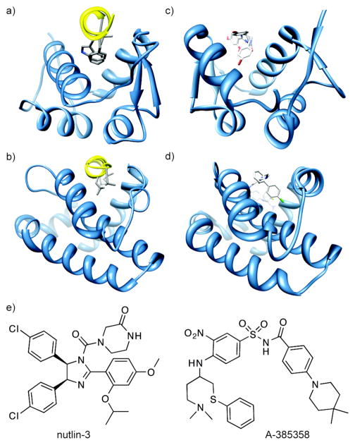
Figure 1.
(a) The p53/HDM2 interaction (PDB code: 1YCR). A helix in the p53 activation domain resides in a deep hydrophobic groove. (b) The pro-apoptotic protein partner Bak bound to the anti-apoptotic protein Bcl-xL (PDB code: 1BXL). (c) Nutlin-3 binds to HDM2 in the same hydrophobic groove occupied by the p53 helix (PDB code: 1rv1). (d) ABT-785358 targets Bcl-xL at the site of its pro-apoptotic binding partners (PDB code: 2o22) (e) The structures of nutlin-3 and A-385358.

