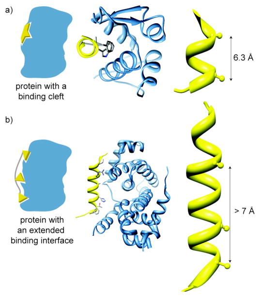
Figure 3.
Helical Interfaces: we have divided helical protein-protein interactions between those that feature clefts for binding (a) and those with extended interfaces (b). The p53/MDM2 (PDB code: 1YCR) (a) and cyclin-dependent kinase6/D-type viral cyclin (PDB code: 1G3N) (b) complexes are representative examples of binding cleft and extended interfaces, respectively. The distance between flanking hot spot residues in the helix of the protein partner of a binding cleft target spans a radius of 7 Å or less (a) and greater than 7 Å but less than 30 Å for an extended interface target.

