
Figure 2. Foxq1sb/sb stomachs are deficient in surface mucin.
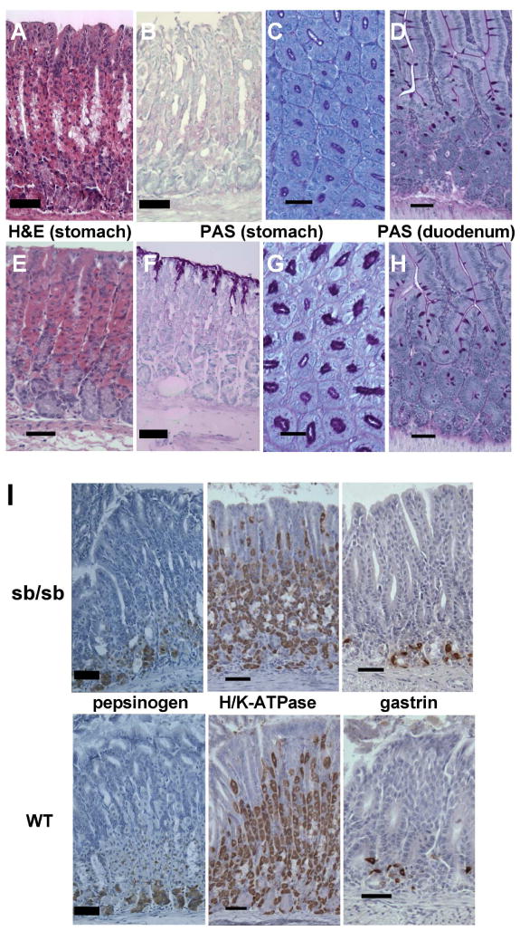
(A–H) Adult mouse stomach or duodenal tissue (Foxq1sb/sb in top 4 panels and controls in lower 4) stained with hematoxylin and eosin (H&E) or periodic acid-Schiff (PAS), as indicated. H&E staining reveals normal character of gastric glandular mucosa; images are taken from the gastric corpus. PAS stain reveals marked attenuation of surface epithelial mucous staining in Foxq1sb/sb stomach (B,C) but normal duodenal goblet-cell staining (D). (I) Immunohistochemistry of adult murine gastric mucosa (Foxq1sb/sb in top 3 panels and wild type in lower 3), showing normal distribution of markers for chief (pepsinogen) and parietal (H/K-ATPase) cells in the corpus and enteroendocrine G (gastrin) cells in the antrum. Scale bars: 60 μm for PAS-stained cross-sections (C,G), 120 μm for all others. Low-power photomicrographs are shown in Suppl. Fig. 1A–D.