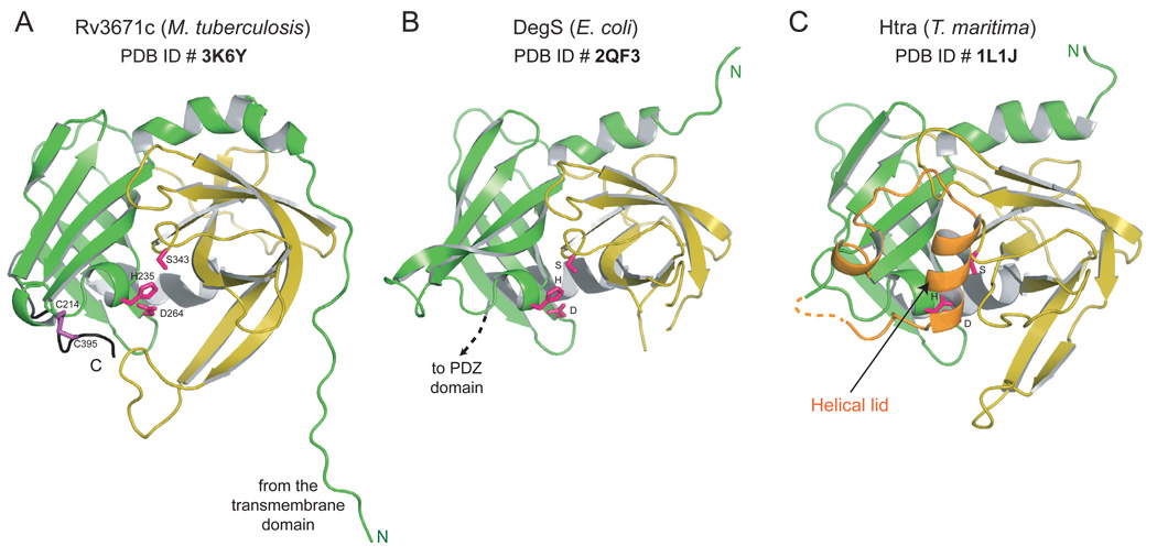
Figure 4.
Structure of Rv3671c. Cartoon representations of A. the structure of Rv3671c B. DegS from E. coli (PDB ID: 2QF3) (Sohn et al., 2007) and C. HtrA from T. maritima (PDB ID: 1L1J) (Kim et al., 2003) are shown in similar orientation. The N- and C-terminal β-barrel subdomains are colored green and yellow, respectively. The catalytic residues and the disulfide bond forming cysteines of Rv3671c are shown as pink and purple sticks.
See also Figure S2.

