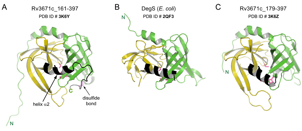
Figure 5.
Cartoon representation of the back views of A. Rv3671c_161-397 (active state) B. DegS from E. coli and C. Rv3671c_179-397 (inactive state) are shown in similar orientations. The kink in helix α2 for Rv3671c_161-397 is indicated by the arrow. Corresponding α2 helices in all panels are colored in black.

