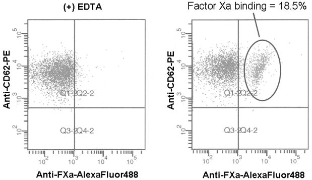
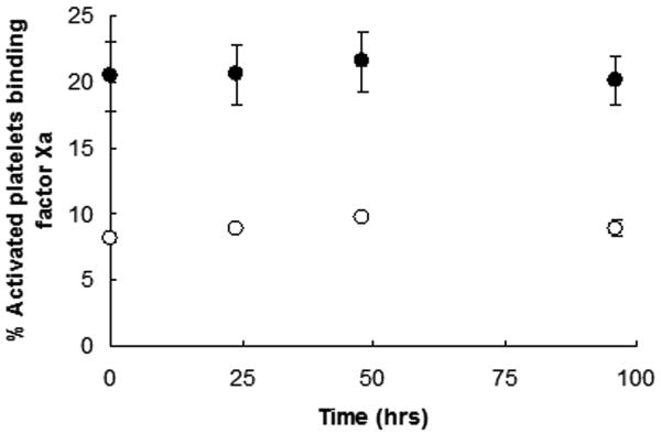
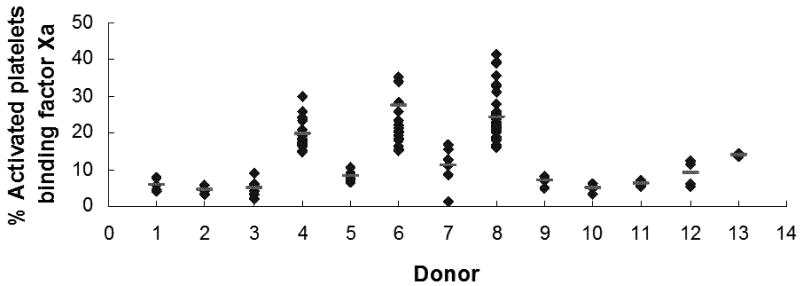
Figure 1. Flow cytomteric assay of factor Xa binding to platelets in whole blood.
(A) Contact pathway-suppressed whole blood was incubated with PAR1 and PAR4 peptides, and factor Va and EGRck-factor Xa at ambient temperature. Platelets were identified using forward scatter and side scatter measurements, and immunostaining with anti-CD61-PerCP (1 nM) (Becton Dickinson, Franklin Lakes, NJ). Activated platelets were identified by immunostaining with anti-P-selectin-PE (0.5 nM) (Becton Dickinson). Factor Xa binding to platelets was determined by immunostaining with anti-factor Xa (FXa)-AlexaFluor488 in the presence or absence of EDTA. Multi-parameter flow cytometric analysis was performed using a BD LSR II Flow Cytometer (Becton Dickinson). The flow cytometry scattergrams shown are representative of those obtained in the presence (left) or absence of EDTA (right). The positive gate was set such that ≤ 2% of the platelets immunostained with the anti-factor Xa antibody in the presence of EDTA were positive. Functional factor Xa binding is reported as the % activated platelets binding factor Xa. (B) Flow cytometric analyses up to 96 hrs subsequent to immunostaining indicated that the percent platelets binding factor Xa was stable when the samples were stored at 4°C. Shown are the results obtained with two individuals. (C) Factor Xa binding to the platelets of 13 healthy individuals was determined repeatedly over a 6 week period as described above. The black diamonds represent single measurements while the light gray horizontal lines indicate the mean of the measurements for that individual.


