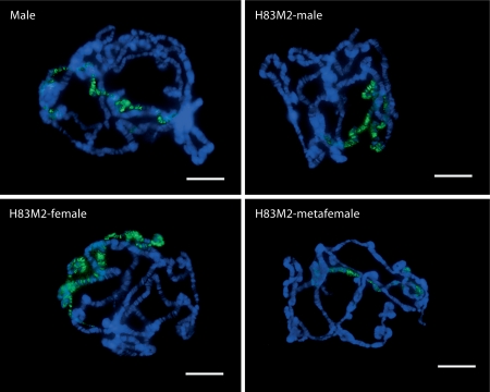
Fig. 3.
Ectopic expression of MSL2 in females and metafemales. Immunofluoresence staining of polytene chromosomes with anti-MSL2 (green) from third instar male, female and metafemale larvae carrying the (w+)H83M2-6I transgene. Nuclei were stained with DAPI in blue. The left upper image represents normal males and upper right the overexpressing MSL2 males. The lower left is ectopically expressing MSL2 females and lower right ectopically expressing MSL2 metafemales. The staining from normal males without (w+)H83M2-6I was used as a comparison. The scale bar represents 15 μm.

