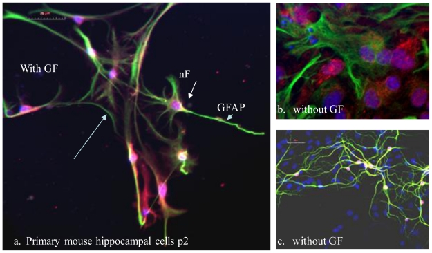
Figure 2.
Primary mouse hippocampal cells cultured in vitro on coverslips with (a) and without growth factors (GF) (b, c), then fixed and stained for neuronal cells with markers neurofilament (nF) red and glial cells with (GFAP) green (a, b), and neural cells MAP2 (c) with DAPI stained nuclei (blue).

