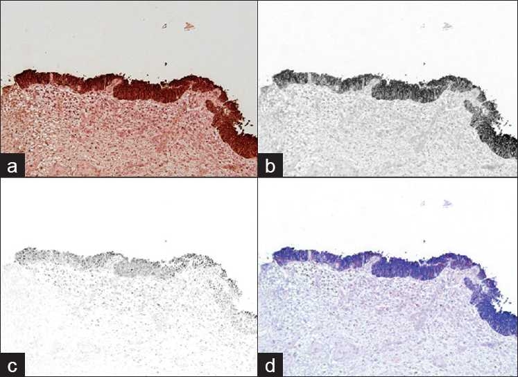
Figure 3.

(a) shows a traditional photographic image (RGB) of the slide. Following “unmixing,” (b) demonstrates CK20 staining the full thickness of the urothelium and (c) shows the P53 staining. (d) shows a composite of the unmixed stains re-assigned with different colors for easier interpretation. This case was graded as 2+ for P53 and 3+ for CK20. The diagnosis given was urothelial carcinoma in situ
