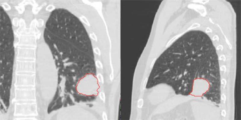
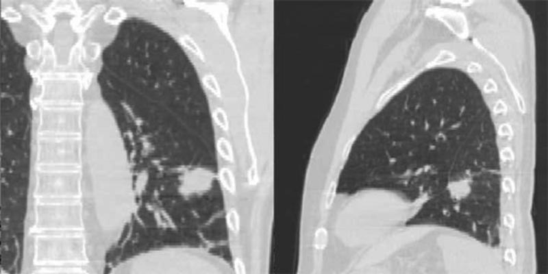
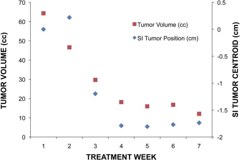
Figure 1.
First (a) and last (b) week breath-hold CT images for Patient 4. Substantial tumor volume regression is evident. GTV from the first week is shown in red. (c) Tumor centroid position relative to bone compared with the tumor volume. As the tumor volume changed, the position of the tumor centroid trended superiorly.


