
Figure 1.
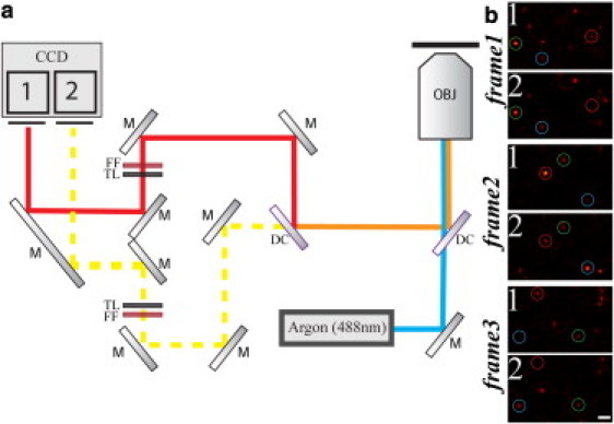
(a) Multicolor nanoscopy setup based on GSDIM, which is essentially a wide-field microscope equipped with a single continuous-wave laser source (Argon 488 nm) and a fast EMCCD camera. Fluorescence is collected by a microscope objective with high numerical aperture (OBJ), separated from the excitation light by a dichroic mirror (DC), split by another dichroic mirror (DC) into two wavelength ranges, and imaged onto two separate areas (area 1 and area 2) of the same EMCCD chip. Labels: (M) Mirror; (FF) fluorescence filter; (TL) tube lens. (b) Three consecutive exemplary camera frames of our GSDIM recordings of Fig. 2 showing on-off blinking of single isolated fluorophores with different ratios of signal counts in the area 1 (>550 nm) and area 2 (<550 nm) of the camera (blue circle, Alexa488 assignment; green circle, Atto532 assignment; red circle, Cy3 assignment). Scale bar 1 μm.