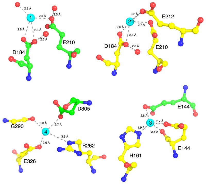
Figure 2.
Interactions of Cadmium with RCK. The crystal structure of RCK in complex with Cd2+ reveals four unique binding sites per RCK dimer. Sites 1 and 2 are in similar location as the Ca2+ binding sites, located on subunits B (green) and A (yellow), respectively. In contrast, Sites 3 and Site 4 are unique to Cd2+. Interacting amino acids residues are shown as ball-and-stick format, with yellow carbons for chain A and green carbons for chain B, red balls for oxygens or water molecules and blue for nitrogen atoms. Cd2+ are shown as cyan spheres. Interaction distances are shown in Å.

