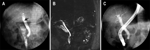
Fig. 1.
(A, B) Endoscopic retrograde cholangiopancreatography (black arrow) and magnetic resonance cholangiopancreatography revealed a 0.5-cm-sized eccentric polypoid lesion at the hilar portion. (C) Follow-up balloon-occluded cholangiography revealed disappearance of the polypoid lesion at the hilar portion.

