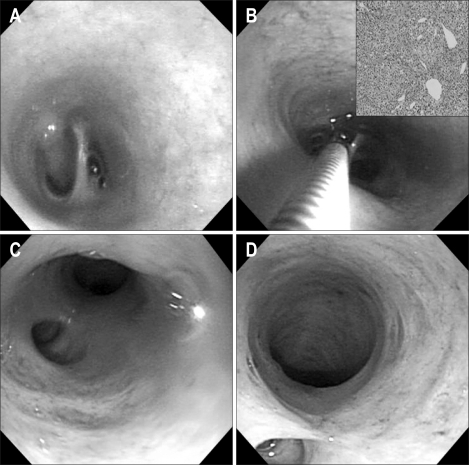
Fig. 2.
(A) Peroral direct cholangioscopic findings demonstrating a 0.5-cm-sized polypoid lesion on the hilar portion. (B) Peroral direct cholangioscopy (PDCS)-guided biopsy specimen of the tiny nodular lesions at the hilar portion. Endoscopic biopsy revealed a hepatocellular carcinoma (H&E stain, ×200; inset). (C, D) Follow-up PDCS with an ultraslim upper endoscope revealed disappearance of the polypoid lesion at the hilar portion.

