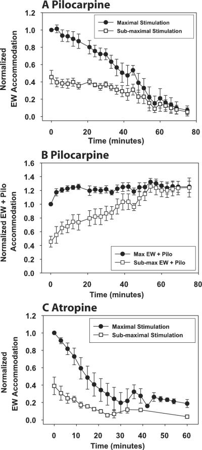
Figure 2.
For all eyes, topical pilocarpine (A) and atropine iontophoresis (C) decreased the EW-stimulated accommodative response amplitudes to submaximal (□) and maximal (●) stimuli. For EW + pilocarpine stimulation (B), responses to maximal EW stimulation increased after the first time point and then remained constant, whereas responses to submaximal EW stimulation increased and approached the maximal responses. Responses are normalized to the pre-pilocarpine maximal EW-stimulated response for each eye. Error bars represent SEM.

