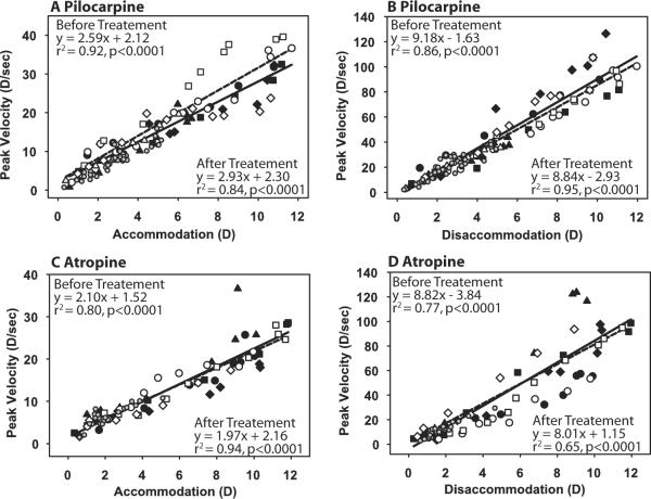
Figure 3.
Main sequence analysis for accommodation (A, C) and disaccommodation (B, D) before (closed symbols, solid lines) and after (open symbols, dashed lines) pilocarpine (A, B) and atropine (C, D). The pre-drug main sequences were determined from responses to stimuli of increasing amplitude. The post-drug main sequences were determined from progressively decreasing responses to the maximal and submaximal stimulus amplitudes. Different symbols represent different monkey eyes. Small gray circles represent the posttreatment response to submaximal stimulus amplitudes for all eyes and are not included in the linear regressions. A significant difference was observed in intercept for the pre-pilocarpine and post-pilocarpine conditions (A; P < 0.05).

