
Figure 5.
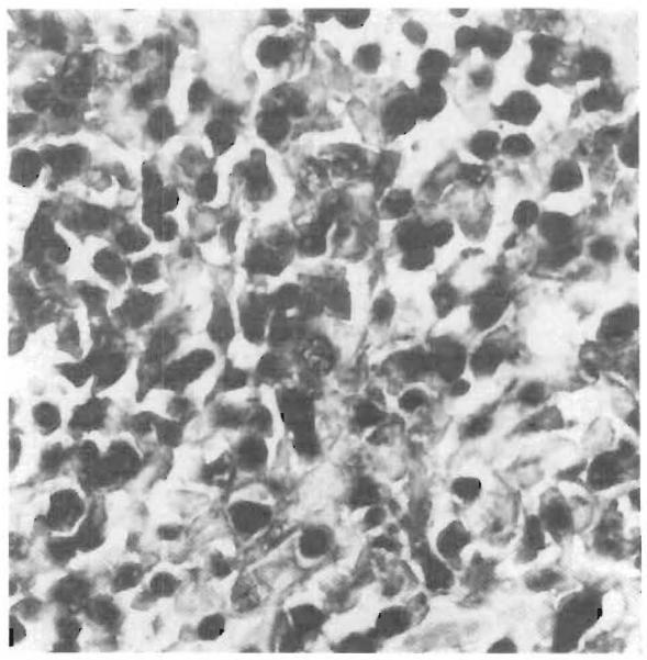
EBV-Infected Lymphocytes in the Lesions of PTLD (×420).
Immunohistochemical evaluation with anti-CD20 antibody, combined with in situ hybridization, showed that in PTLD the majority of EBV-infected lymphocytes are B cells. Cells positive for CD20 are stained brown, and those positive for EBER-1 mRNA are stained blue-black.