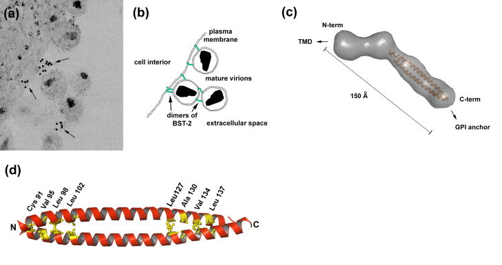
Figure 2. Mechanism of restricted virion release by BST-2.
(a) Immuno-electron microscopic data indicate that BST-2 is appropriately positioned to directly tether mature virions to the plasma membrane and is incorporated into virions. The cell cytoplasm is at the left; the extracellular space is at the right and includes mature virions. Arrows indicate immuno-label for the BST-2 ectodomain. Reprinted with permission from Ref. [31]. (b) Model in which BST-2 dimers (green) embed one end in the plasma membrane and the other in the virion membrane, retaining virions on the cell surface. BST-2 dimers may also link virions to each other. An alternative model is discussed in the text. (c) Structure of the dimeric BST-2 ectodomain obtained by small angle X-ray scattering (molecular surface in gray) and X-ray crystallography (modified from Ref. [21]). The parallel, dimeric coiled-coil is colored orange. Abbreviations: Å, angstroms. (d) Crystallographic structure of the dimeric coiled-coil indicating residues important for the restriction of virion release (PDB accession code: 2x7a; structure drawn using PyMOL).

