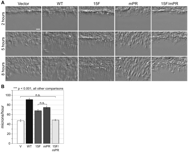
Figure 2. p130Cas SD and SBD signaling stimulate wound healing migration.
Monolayer scratch wounds were generated and cells were imaged over 8 hours by live DIC microscopy. (A) Representative images of cells expressing either p130Cas WT, 15F, mPR, or 15F/mPR variants at 2, 5, and 8 hours post wounding. Vector only control cells are also shown. Scale bar is 50 µm. (B) Migration rates were quantified by tracking nuclei of 75 individual cells (25 each from 3 separate wounds) for each cell type. Mean migration rate was calculated as the total distance traveled over the 2 to 8 hour period after wounding. Bars indicate s.e.m. Significance values were determined by one-way ANOVA followed by Tukey-Kramer post hoc testing; n.s. (not significant), ***p<0.001.

