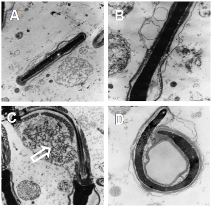
Figure 2. Ultrastructural changes.
Transmission-electron micrographs of rabbit sperm heads from NCR (A) and HCR (B to D). Notice the small vesicles in the acrosome region in B and the long side fold of sperm head in D. Some sperm cells show the remaining residual body (white empty arrow, C). A and C, X 12,000; B and D, X 20,000.

