
Figure 5.
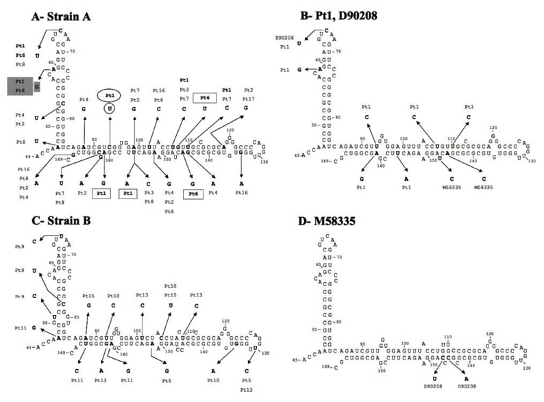
Prediction of stem-loop V and VI secondary structures. Numbering starts at the A of the AUG initiation codon. Variable positions are indicated in bold. (A) Predicted secondary RNA structure of the consensus strain A sequence. Differences relative to the consensus sequence observed in the patients who seroconverted (patients 1 and 6) are indicated by the bold patient’s number. The circled U was found in the major quasispecies variant of patient 1. Positions highlighted in gray correspond to substitutions found in both patients 1 and 6. Squared differences relative to the consensus sequence were found either in patient 1 or in patient 6. (B) Predicted secondary RNA structure of patient 1’s major quasispecies sequence, which was identical to that of reference strain D90208. (C) Predicted secondary RNA structure of the consensus of strain B sequences. (D) Predicted secondary RNA structure of reference strain M58335.