
Figure 3. Polarized secretion of cytokines and growth factors by mouse liver cyst epithelial cells.
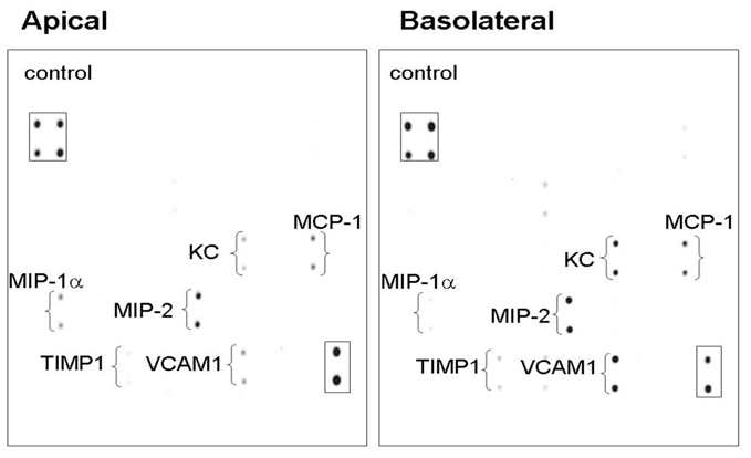
Primary cultures of isolated liver cyst epithelial cells from pkd2(WS25/−) mice grown on semi-permeable supports form high resistance monolayers (Rt>1,000 Ω·cm2). Cytokine array analysis of media collected after 72 hours of incubation showed a parallel pattern of cytokine and growth factor release into both the apical and basal media. Boxes show internal array controls.