
Figure 5. IL-8 and liver cyst fluid promote Mz-ChA1 cell proliferation.
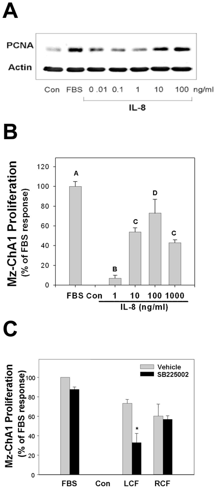
Treatment of Mz-ChA1 cells with IL-8 for 24 hrs induced cell proliferation. (A) Western blot analysis of PCNA showed Mz-ChA1 cells had modest responses with 0.01 to 1.0 ng/ml IL-8 and marked proliferative responses when treated with 10 to 100 ng/ml IL-8. Actin served as a loading control. Blot is representative of three separate experiments. (B) Alamar Blue reduction assays showed a modest proliferative response from 1 ng/ml IL-8 and an apparent maximal response at 100 ng/ml. From an analysis of variance, bars without a shared letter (e.g. A, B, C, D) are significantly different from each other. (C) Stimulation with 10% human liver cyst fluid markedly induced proliferation of unsupplemented Mz-ChA1 cells. This response was significantly inhibited by pretreatment with 50 nM SB225002 (*p <0.05 vehicle vs SB225002). Addition of 10% renal cyst fluid also induced the proliferation of Mz-ChA1 cells. This response, however, was not inhibited by pretreatment with SB225002.