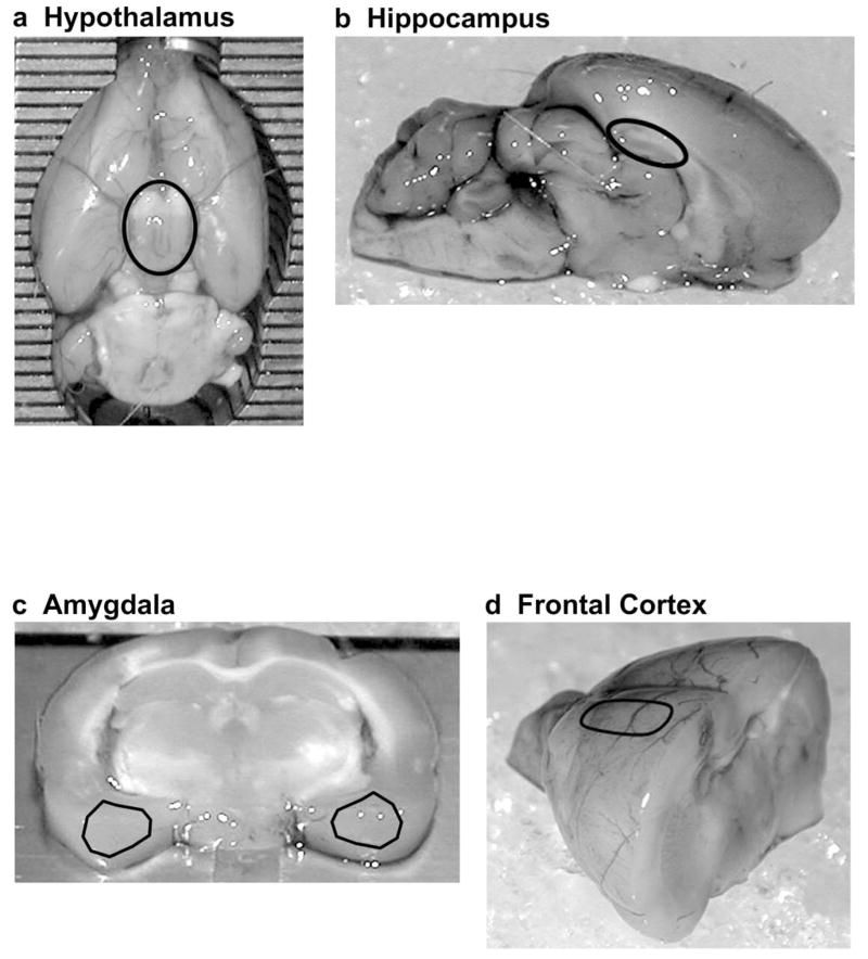
Fig 1.
Regions of the brain used for assessing mRNA levels. (a) The hypothalamus (black circle) was extracted from the ventral surface of the rat brain. (b) The hippocampi (black circle) were dissected from a mid-sagittal section of the rat brain. (c) 2.0 mm thick sections of amygdalae (black triangles) were dissected from a coronal section of the rat brain. (d) Frontal cortices (black oval) were dissected from an anterior portion of the rat brain. Dissection techniques and approximate stereotaxic coordinates for these brain regions are described in greater detail under “Brain Dissection” in the Methods.

