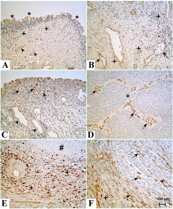
Figure 4.
Immunohistochemical staining of SPARC in normal human bladder, cystitis, invasive and noninvasive urothelial carcinoma. (A). Localization of SPARC in normal human bladder tissue sample. * indicates SPARC staining in the normal urothelium. + indicates staining in the stromal cells. Arrow heads indicate lack of staining in blood vessels. (B). Localization of SPARC in a bladder tissue sample obtained from a patient with cystitis. + indicates frequent staining of stromal cells. Arrows indicate moderate staining in blood vessels. (C). Localization of SPARC in blood vessels of bladder tissue obtained from a patient with cystitis. Arrows indicate strong staining in the newly formed blood vessels, whereas mature blood vessels in deep lamina propria are weakly positive or negative for SPARC (arrow heads). (D). Localization of SPARC in low grade urothelial papillary carcinoma. The tumor cells did not stain for SPARC whereas few stromal cells in the papillary core (+) stained for SPARC. Arrow indicates strong staining in small blood vessels. (E). Localization of SPARC in low grade urothelial papillary carcinoma containing areas of necrosis. # indicates an area of necrosis, around which a large number of SPARC positive (+) stromal cells are present. (F). Localization of SPARC in high grade invasive bladder cancer. The tumor cells did not stain for SPARC whereas the desmoplastic stromal cells both in and around the invasive carcinoma stained strongly (+). Arrows indicate positive staining of the endothelial cells of the blood vessels. All images are at a magnification of X200. Bar = 100 μm.

