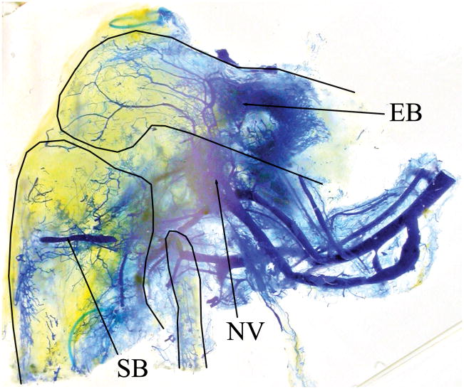
Figure 5.
Optically cleared knee allotransplant (right side, from medial). Thin solid lines show the outlines of the femur, tibia and fibula. Blue endovascular polymer visualizes capillary networks from three locations: saphenous (SB) and epigastric (EB) a/v bundles and the transplant nutrient vessels (NV). (Refer to Figure 2 for orientation.)

