Figure 6.

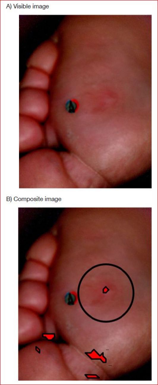
(A) Visible image of plantar ulcer without ulceration 116 days earlier. (B) Composite image produced using Method 2 with red overlays identifying areas at risk of ulceration. Circle identifies where the ulcer eventually formed.

(A) Visible image of plantar ulcer without ulceration 116 days earlier. (B) Composite image produced using Method 2 with red overlays identifying areas at risk of ulceration. Circle identifies where the ulcer eventually formed.