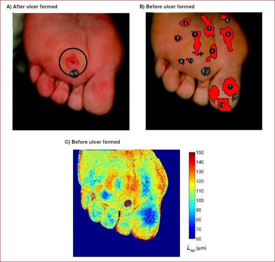
Figure 8.

(A) Plantar and toes of the right foot exhibiting a newly formed ulcer (July 27, 2007). (B) Composite image where the red overlays indicate regions at risk of ulceration56 (July 10, 2007). (C) Epidermal thickness retrieved from images taken before ulceration showing thickening around the site where ulcer eventually formed.
