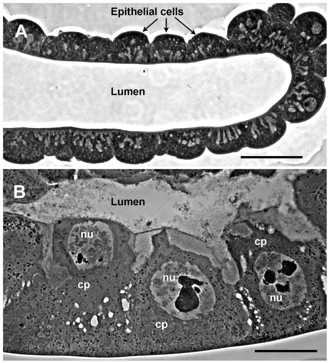
Fig. 1.
Light micrographs of thick sections through (A) healthy and (B) MdSGHV-infected salivary glands. Images were taken at identical magnification with bars equal to 50 μm. The salivary gland supports viral replication along its entire length. The relative number of cells in healthy and infected glands remains constant; the enlarged state of the infected gland is due to extensive cellular and nuclear hypertrophy induced by viral replication. Cp, cytoplasm; nu, nucleus.

