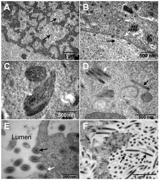
Fig. 2.
Transmission electron micrographs of hypertrophied salivary glands. (A) The enlarged nuclei were characterized by virogenic stroma within which the nucleocapsids (back arrows) were assembled and then (B) translocated to the nuclear membrane where they were observed to exit (black arrow) the nucleus via the nuclear pores and to be released (white arrow) into the region of the perinuclear cistern. (C) Virus particles (white arrow) appear to be associated with endoplasmic reticulum. (D) Both clusters (white arrow) and individual (black arrow) enveloped virions are found throughout the cytoplasm. (E) The enveloped virions (black arrow) migrate preferentially to the cell membrane (white arrow) and are released into the gland lumen via a budding process. (F) As the infection progresses the lumen becomes filled with enveloped virions (black arrows).

