
Fig. 1.
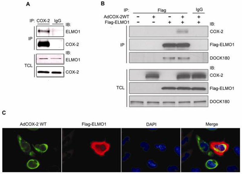
ELMO1 forms a complex with COX-2 in mesangial cells. (A) HMC lysates were immunoprecipitated with either COX-2 antibody, or control IgG, and co-precipitation of endogenous ELMO1 was determined by immunoblotting. Expression of the endogenous proteins was determined by immunoblotting of TCL. (B) Flag-ELMO1 was transfected into THMC, and co-precipitation of infected COX-2 and endogenous DOCK180 were determined by immunoblotting. Expression of the endogenous proteins or overexpressed proteins was determined by immunoblotting of TCL. (C) Flag-ELMO1 and AdWTCOX-2 expressing THMC were stained with anti-Flag (red) and anti-COX-2 (green), and their chromatin was stained with DAPI (blue). Colocalization of ELMO1 and COX-2 appears in yellow in the merge panel. Data are representative of three independent experiments.