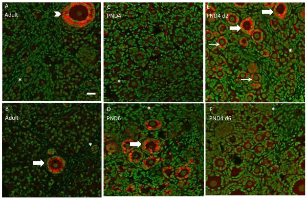
Figure 1.
Ovarian AMH protein staining. In vivo ovaries were collected from adult F344 rats (16 weeks; A and B), PND4 (C) or PND6 (D) rats. PND4 ovaries were cultured in vitro with control medium for 2 (E) or 6 days (F). All ovaries were processed for confocal microscopy as described in materials and methods. All panels are a combined overlay of AMH (Cy-5 red stain) and genomic DNA (green YOYO stain) at 40X magnification. Thin arrow, small primary follicle; thick arrow, large primary follicle; arrowhead, secondary follicle; asterisk, primordial follicle. Scale-bar equal to 25 μM.

