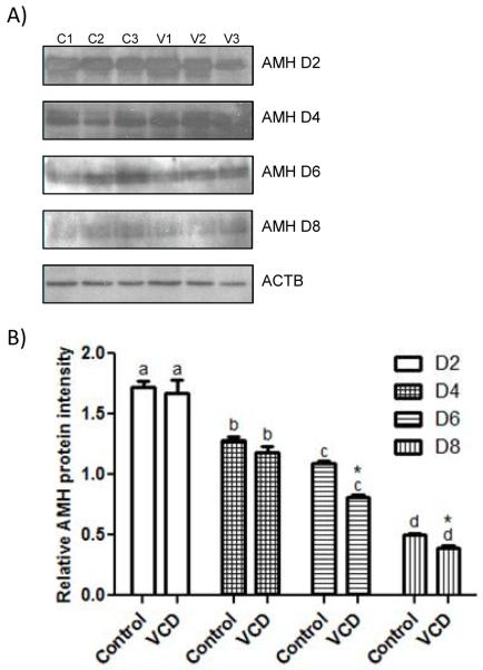
Figure 3.
Effect of VCD on expression of AMH protein. Ovaries from PND4 F344 rats were cultured with control medium ± VCD (30 μM) for 2-8 days. (labeled D2-8) (A) Representative Western blots for AMH and β-actin (ACTB) expression in cultured ovaries in control medium (C1, C2, C3) or medium containing VCD (V1, V2, V3). One representative ACTB blot from d2 is shown, however, ACTB expression was measured at each time point. (B) AMH protein intensity was normalized to ACTB protein and expressed as mean ± SEM; n = 3; 10 ovaries/pool for cultured ovary samples; different superscript letters indicate a differences between time points (P < 0.05) difference; *P < 0.05, different from control.

