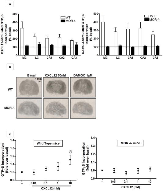
Figure 1. CXCR4 coupling to G proteins is reduced in the brains of MOR−/− animals.
(a) Coronal brain sections of wild type and MOR−/− mice were treated with CXCL12 (50nM) or DAMGO (1μM) and processed for GTPγS autoradiography. Analysis was performed in different areas of the brain: the medial cortex (MC), lateral cortex (LC), and hippocampus (fields CA1, CA2 and CA3). Data are expressed as mean ± SEM of n=3 animals per group. A statistically significant difference was observed in MOR−/− compared to WT brain sections in all of these areas (CXCL12-induced GTPγS incorporation: t8=4.159, P=0.0032; DAMGO-induced GTPγS incorporation: t8= 7.984, P < 0.0001). (b) Representative autoradiographs of GTPγS incorporation after vehicle-(i.e. basal), CXCL12- (center panels), and DAMGO- (right panels) stimulation of WT and MOR−/− pups are shown. Scale bar, 1mm. (c) Brain homogenates (cortices and hippocampi) of P2-P11 WT (left panel) and MOR−/− (right panel) pups were exposed to different doses of CXCL12, as indicated. Graphs represent the mean ratio of agonist-stimulated GTPγS binding over basal (n=5) ± SEM. [F4, 95= 2.876, O P > 0.05 versus basal].

