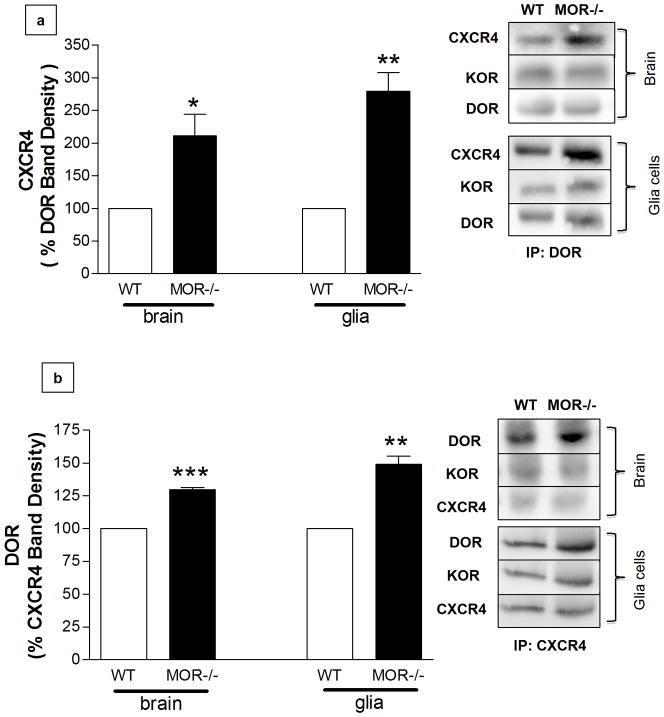
Figure 5. Increased co-immunoprecipitation (IP) of CXCR4 with DOR in brain tissue and glial cultures.
(a) After blotting for CXCR4 and KOR, membranes were stripped and reprobed for DOR to confirm equal loading (IP in brain homogenates: t6= 3.387, * P < 0.01, Student’s t-test, n=4 animals/group; IP in glia cultures: t6= 6.274, ** P < 0.005, Student’s t-test, n=4 glial cultures/group). (b) After blotting for DOR and KOR, membranes were stripped and reprobed for CXCR4 to confirm equal loading (IP in brain homogenates: t4= 19.35, ***P < 0.0001, Student’s t-test, n=3 animals/group; IP in glia cultures: t4= 7.771, *P < 0.005, Student’s t-test, n=3 glial cultures/group).

