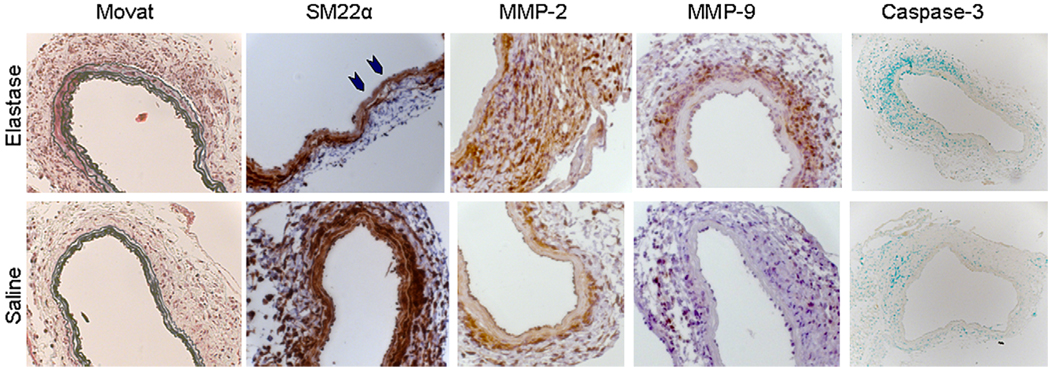
FIGURE 3.
Histochemistry and immunohistochemistry in mice 7 days after elastase or saline perfusion. Little differences in elastin are evident by means of Movat staining, with intact elastin in both treatment groups. Some early degradation might be present in regions of the aorta (blue arrowheads) after elastase perfusion compared with that seen after saline perfusion. Expression of matrix metalloproteinase (MMP) 2 and 9 appears to be upregulated at 7 days after elastase compared with that seen after saline. Minimal activated (cleaved) caspase-3 staining is present in either group (brown positive stain, methyl green nuclear counterstain). Images were obtained at 25× magnification, except for caspase-3, which was obtained at 10× magnification.

