
Figure 5. EMG recordings in 3 different spontaneously breathing rats.
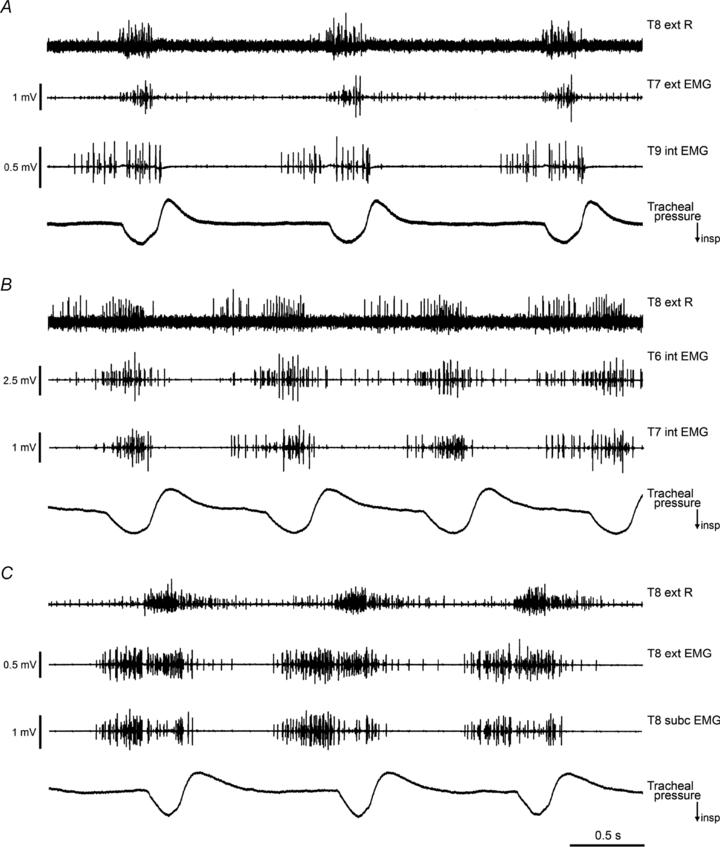
In each animal, the efferent discharges from the T8 external intercostal nerve on the right side are shown (T8 ext R), together with two EMG recordings on the left, from the surface of the external (ext) or internal (int) intercostal muscle layers or subcostalis (subc), as indicated. The tracheal pressure record indicates the phase of respiration, inspiration downwards. The intercostal nerves and muscles for all segments on the left were intact when these records were taken. Levator costae muscles had been removed from T7–T10 for A and from T6–T10 for B and C. Time calibration applies to all 3 panels. A, decerebrate following halothane, 2 h 25 min after decerebration. B, α-chloralose following halothane, 4 h 45 min after switching to α-chloralose. C, decerebrate following halothane, 2 h 50 min after decerebration.