
Figure 6. Effects of nerve section on EMG recordings in one intercostal space.
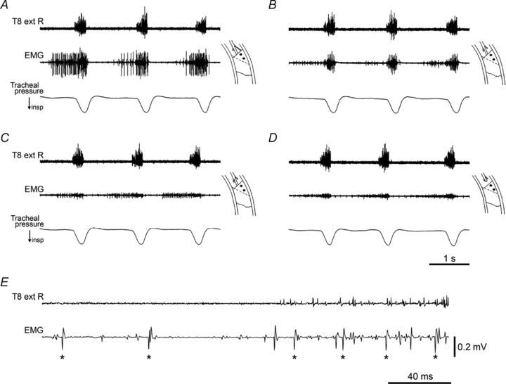
A–D, EMG recordings at one site shown in time order, with successive nerve or muscle sections. Diagrams indicate the recording site, at the proximal border of the left internal intercostal muscle in T10. In this and subsequent figures, the black dots indicate the positions of the two recording wires. Top trace is the recording from the external intercostal nerve of T8 on the right side. A, the proximal part of the external intercostal muscle in T10 and its nerve had been removed (dotted line in diagrams indicate its original proximal border), T9 had been denervated and the external intercostal nerve cut in T11. B, following section of the internal intercostal nerve filament. C, following additional removal of levator costae muscle in T11. D, following additional section of the internal intercostal nerve in T10. See text for more details. E, extract from the nerve and EMG recordings in A (start of 2nd inspiration), shown on an expanded time scale. Large EMG spikes (asterisks) show a probable single motor unit that was active in both inspiration and expiration. Time calibration in D applies to A–D. Voltage calibration in E applies to A–E. α-Chloralose following halothane.