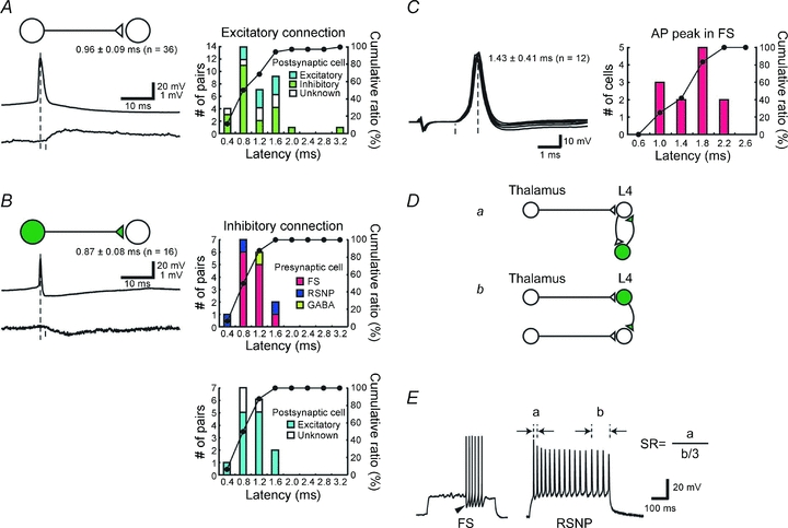
Figure 2. Properties of monosynaptic intracortical connections obtained by dual whole cell recordings from neurons located in a single barrel.

A and B, latency analysis of monosynaptic excitatory (A) and inhibitory (B) connections. Distribution histograms and cumulative plots are shown on the right. Postsynaptic cell types, either excitatory or inhibitory, are indicated by colour in A. In B, the same histograms are presented with different breakdown of presynaptic (upper) and postsynaptic (lower) cell types. C, histogram showing latencies for action potentials measured from the onsets of EPSPs to the action potential peaks in FS cells. D, schematic drawings for generating fast IPSPs. a, thalamic activation of excitatory cells produces feedback IPSPs via neighbouring inhibitory cells, and thus IPSPs are trisynaptic from the thalamic cell. b, direct activations of inhibitory cells from the thalamus that are fed forward to excitatory cells. Thus IPSPs are disynaptic from the thalamus, and therefore faster by one synapse than the trisynaptic IPSP shown in a. E, example responses of inhibitory neurons to depolarizing current injections. GFP-positive neurons were divided into fast spiking (FS) or regular spiking non-pyramidal (RSNP) by the spike frequency adaptation ratio (SR) and large monophasic AHP (arrowhead) and half-height spike width. SR was defined as the ratio of the first interspike interval divided by the average of the last three interspike intervals, as shown here.
