
Figure 5. White matter stimulation also caused latency differences.
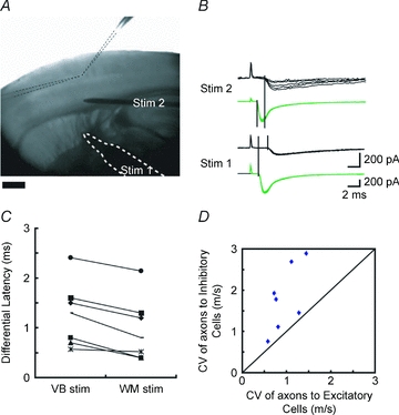
A, photomicrograph showing the experimental set-up. Two recording electrodes (delineated by dotted lines) were inserted into a single barrel, a stimulating electrode (bordered by white dotted line, Stim. 1) was placed on the thalamic fibres issuing from ventrobasal nucleus of the thalamus, and an additional stimulating electrode was placed a little above the white matter (Stim. 2). B, sample EPSCs recorded from GFP-positive (green) and -negative (black) cells in response to the stimulation to Stim. 1 and Stim. 2. Holding potential =−70 mV. No bicuculline was added. C, differences in latencies of EPSCs between excitatory and inhibitory cells were plotted at two stimulation sites, showing that there were still latency differences even when the stimulation was applied to the white matter (Stim. 2), but the differences became smaller. D, from the differences in latencies and the distances between two stimulation sites, conduction velocities (CV) for thalamic axons innervating excitatory and inhibitory cells could be calculated. CVs of axons to inhibitory cells were plotted against those of axons to excitatory cells in simultaneous recordings of 7 pairs. All of the points fell above the diagonal line, indicating that CVs for axons innervating inhibitory cells were faster than those for excitatory cells between VB and white matter.