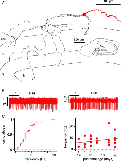
Figure 2. Basic anatomical and physiological properties of Cajal–Retzius cells.

A, reconstruction of a biocytin-filled Cajal–Retzius cell. Notice the single dendrite (red) and axon (black) emerging form opposite sides of the soma. Notice also the axonal targeting of stratum lacunosum-moleculare and of the dentate gyrus. l-m, stratum lacunosum-moleculare; m, stratum moleculare; g, stratum granulosum; h, hilus. Inset shows the entire slice. B, spontaneous firing of Cajal–Retzius cells in the absence of glutamatergic and GABAergic ionotropic transmission. Cell-attached recordings showing action currents in neurons from slices at different postnatal developmental stage. C, summary plots showing the cumulative distribution of spontaneous frequency (left) and the lack of correlation between spontaneous frequency and postnatal age of the animals used in the experiments. See also Supplemental Fig. S1.
