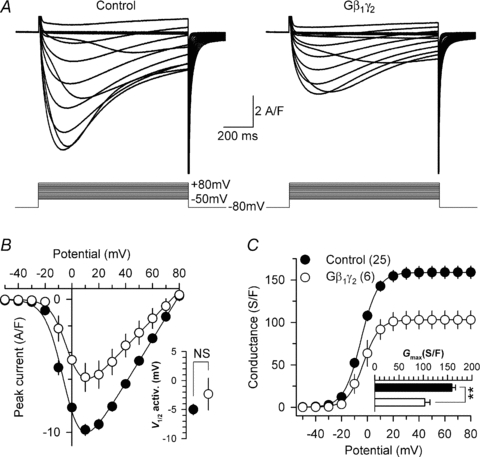
Figure 2. Expression of G-protein β1γ2 dimers reduces L-type calcium current density in skeletal muscle fibres.

A, representative sets of Ca2+ current traces recorded from a control (left panel) and from a Gβ1γ2 dimer-expressing fibre (right panel) in response to 1 s depolarizing steps to values ranging between −50 mV and +80 mV from a holding potential of −80 mV. B, corresponding mean voltage dependence of the peak Ca2+ current density. Inset presents the mean half-maximal activation potential for control and Gβ1γ2-expressing fibres. C, corresponding mean values for the voltage dependence of Ca2+ conductance in the two populations. Inset presents the mean maximal conductance for control and Gβ1γ2-expressing fibres. The maximal conductance was reduced by 35% (P= 0.001) in the Gβ1γ2-expressing fibres.
