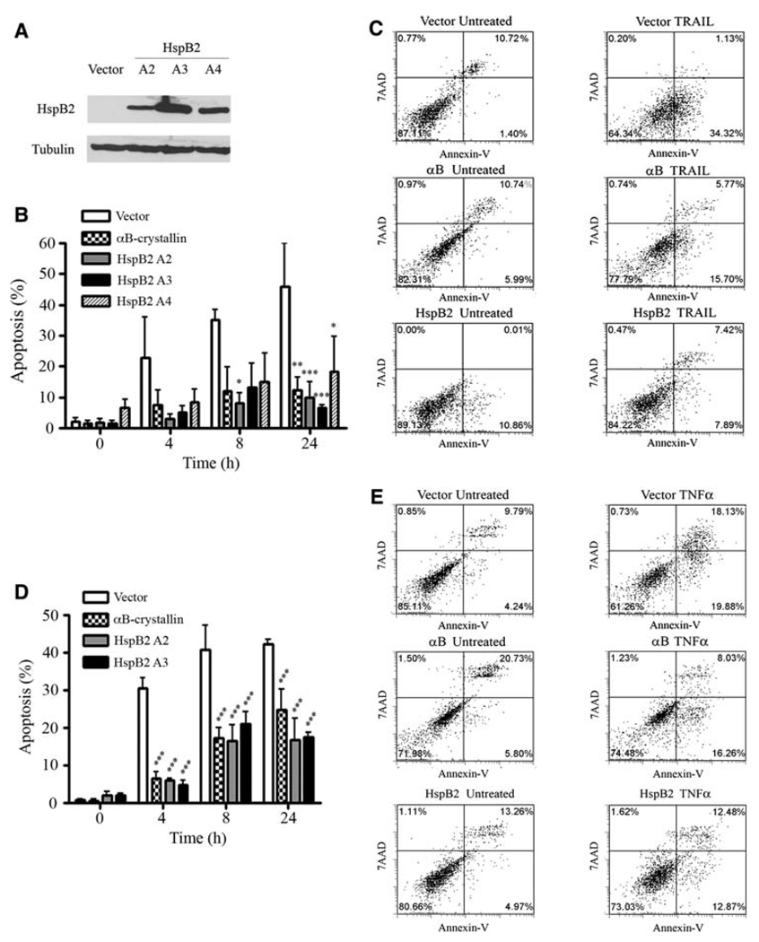
Fig. 2.
HspB2 inhibits TRAIL and TNF-α-induced apoptosis. a Immunoblot of MDA-MB-231 cells stably transfected with FLAG-tagged wild-type HspB2 cDNA or empty vector. The ectopically expressed proteins were detected using the FLAG M2 monoclonal antibody. b MDA-MB-231 breast cancer cells stably expressing HspB2 (three clones, A2, A3, and A4), αB-crystallin or empty vector were treated with 500 ng/ml TRAIL for 0–24 h, and the percentage of cells with apoptotic nuclei were scored. The data represent the mean ± SEM of three independent experiments (*P < 0.05, **P < 0.01, ***P < 0.001 versus vector control for each time point). c MDA-MB-231 stably expressing HspB2, αB-crystallin or empty vector were untreated or treated with 500 ng/ml TRAIL for 4 h. After 4 h, cells were analyzed by flow cytometry for Annexin V-PE and 7AAD fluorescence. The percentage of cells in each quadrant is indicated. d MDA-MB-231 breast cancer cells stably expressing HspB2, αB-crystallin or empty vector were treated with 10 ng/ml TNF-α and 1 µg/ml cycloheximide for 0–24 h, and the percentage of cells with apoptotic nuclei was scored. The data represent the mean ± SEM of three independent experiments (***P < 0.001 versus control at each time point). e MDA-MB-231 cells stably expressing HspB2, αB-crystallin or empty vector were untreated or treated with 10 ng/ml TNF-α and 1 µg/ml cycloheximide for 4 h, at which time cells were analyzed for Annexin V-PE and 7AAD fluorescence by flow cytometry

