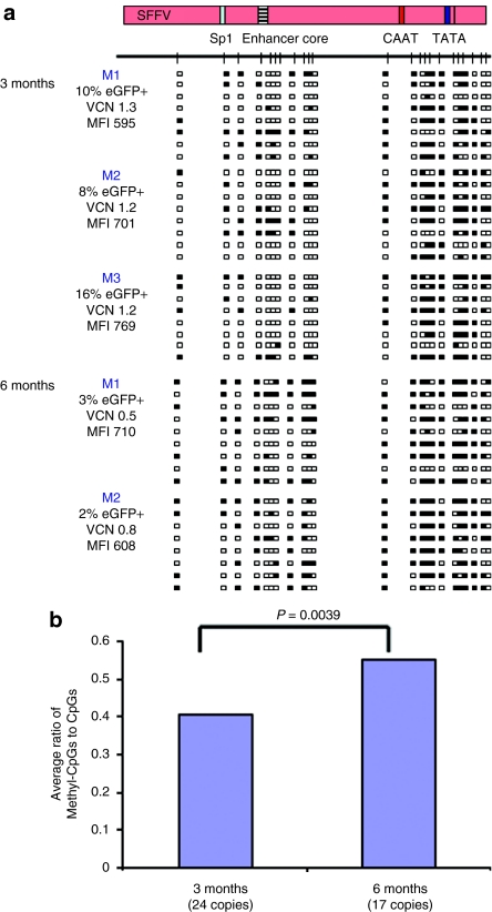
Figure 4.
DNA methylation analysis of the SFFV promoter in vivo following gene transfer to mouse hematopoietic stem cells. Mouse bone marrow lin-selected stem cells were transduced with SIN-LV SFFV-eGFP virus and engrafted into lethally irradiated animals. Bone marrow from transduced mice was isolated at 3 (n = 3) and 6 (n = 2) months after transduction and subjected to DNA methylation analysis. (a) DNA methylation analysis of SFFV proviral sequences from five transduced mice. Note: the SFFV promoter is densely methylated in all five mice. Methylated CpG sites were concentrated around the CAAT/TATA box core promoter elements in contrast to the Sp1/enhancer core region. MFI, mean fluorescence intensity; VCN, vector copies per cell. (b) Comparison of the average ratio of methylated: nonmethylated CpG dinucleotides among vector copies analyzed at 3 months and 6 months after transduction. Methylated CpG sites increased significantly over this time period (The P values were determined using the Wilcoxon rank-sum test).

